The Reproductive System of Flowers
Flowers are not only captivating in their beauty and fragrance but also in their intricate reproductive systems. These exquisite creations of nature possess a remarkable array of structures and mechanisms that allow them to reproduce and ensure the continuation of their species. In this blog post, we will explore the fascinating world of the reproductive system of flowers and unravel the secrets behind their successful propagation.
The Anatomy of Flower Reproduction:
A flower's reproductive system comprises both male and female reproductive organs, each playing a crucial role in the process of pollination and fertilization. Let's delve into the key components:
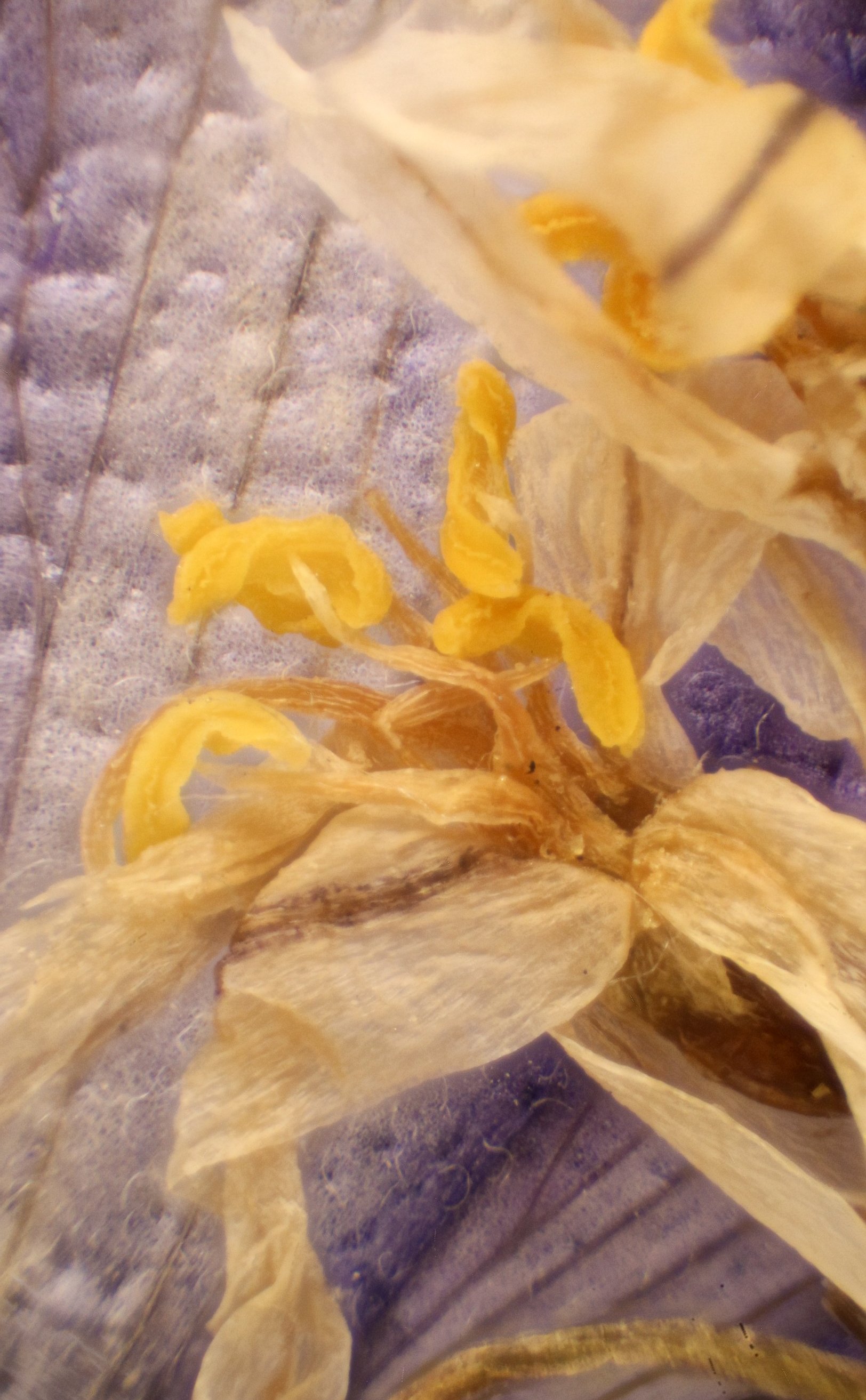
The Stamen - Male Reproductive Organ: The stamen is the male reproductive organ of a flower, consisting of two main parts: the anther and the filament. The anther, situated at the tip of the filament, contains pollen sacs called microsporangia, which produce pollen grains. These pollen grains carry the male gametes, or sperm cells, necessary for fertilization.
The Pistil - Female Reproductive Organ: The pistil, also known as the carpel, is the female reproductive organ of a flower. It consists of three main parts: the stigma, style, and ovary. The stigma, located at the top of the pistil, is a sticky platform that captures pollen grains. The style is a slender tube that connects the stigma to the ovary. Finally, the ovary is a swollen base containing one or more ovules.
Pollination - The Transfer of Pollen: Pollination is the process of transferring pollen grains from the anther to the stigma. It can occur through various mechanisms, including wind, water, or animal pollinators like insects, birds, or mammals. Insects, particularly bees, are among the most common and efficient pollinators, attracted by the flower's color, scent, and nectar. When a pollinator visits a flower, pollen grains may stick to its body or get deposited on the sticky surface of the stigma.
Fertilization - Fusion of Gametes: Once a pollen grain reaches the stigma, it germinates and develops a pollen tube that grows down the style towards the ovary. The pollen tube delivers the male gametes to the ovule, located within the ovary. Fertilization occurs when a sperm cell fuses with an egg cell inside the ovule, leading to the formation of a zygote. This fertilized ovule develops into a seed.
Fruit Development and Seed Dispersal: After fertilization, the ovary undergoes remarkable changes, transforming into a fruit that protects the developing seeds. Fruits come in various shapes, sizes, and textures, enticing animals to eat them and disperse the enclosed seeds. This dispersal mechanism ensures the wider distribution of seeds, increasing the chances of successful germination and the growth of new plants.
The Diversity of Floral Reproductive Strategies:
Flowers exhibit an astonishing diversity of reproductive strategies, reflecting the adaptability of different plant species to their environments. Some flowers have both male and female reproductive organs in the same flower, known as "perfect" or "hermaphroditic" flowers. Others have separate male and female flowers on the same plant, called "monoecious" plants, or on different plants, known as "dioecious" plants.
Additionally, some flowers employ strategies to prevent self-fertilization, promoting outcrossing and genetic diversity. These mechanisms include self-incompatibility, where a flower rejects its own pollen, and dichogamy, where the male and female reproductive organs mature at different times to avoid self-pollination.
The reproductive system of flowers is an astonishing testament to nature's ingenuity. Through the interplay of male and female reproductive organs, the processes of pollination, fertilization, and seed dispersal, flowers ensure their survival and perpetuation. As we marvel at the beauty and diversity of flowers around us, let us appreciate the incredible mechanisms that underpin their reproductive success and the vital role they play in sustaining our ecosystems.
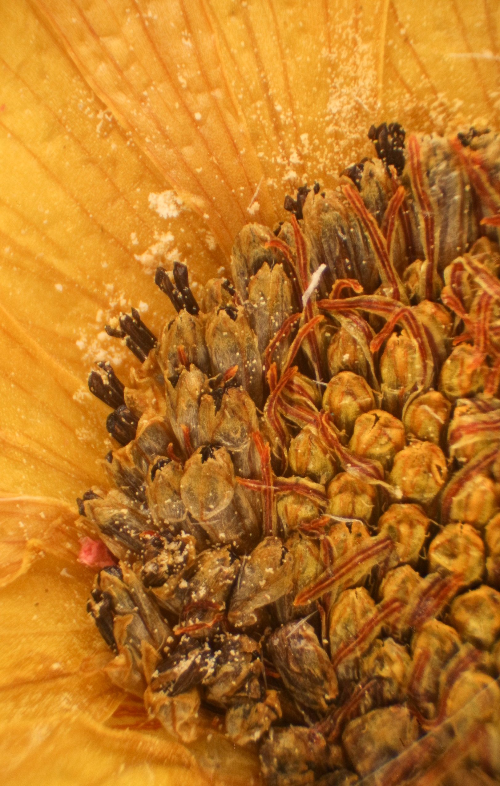
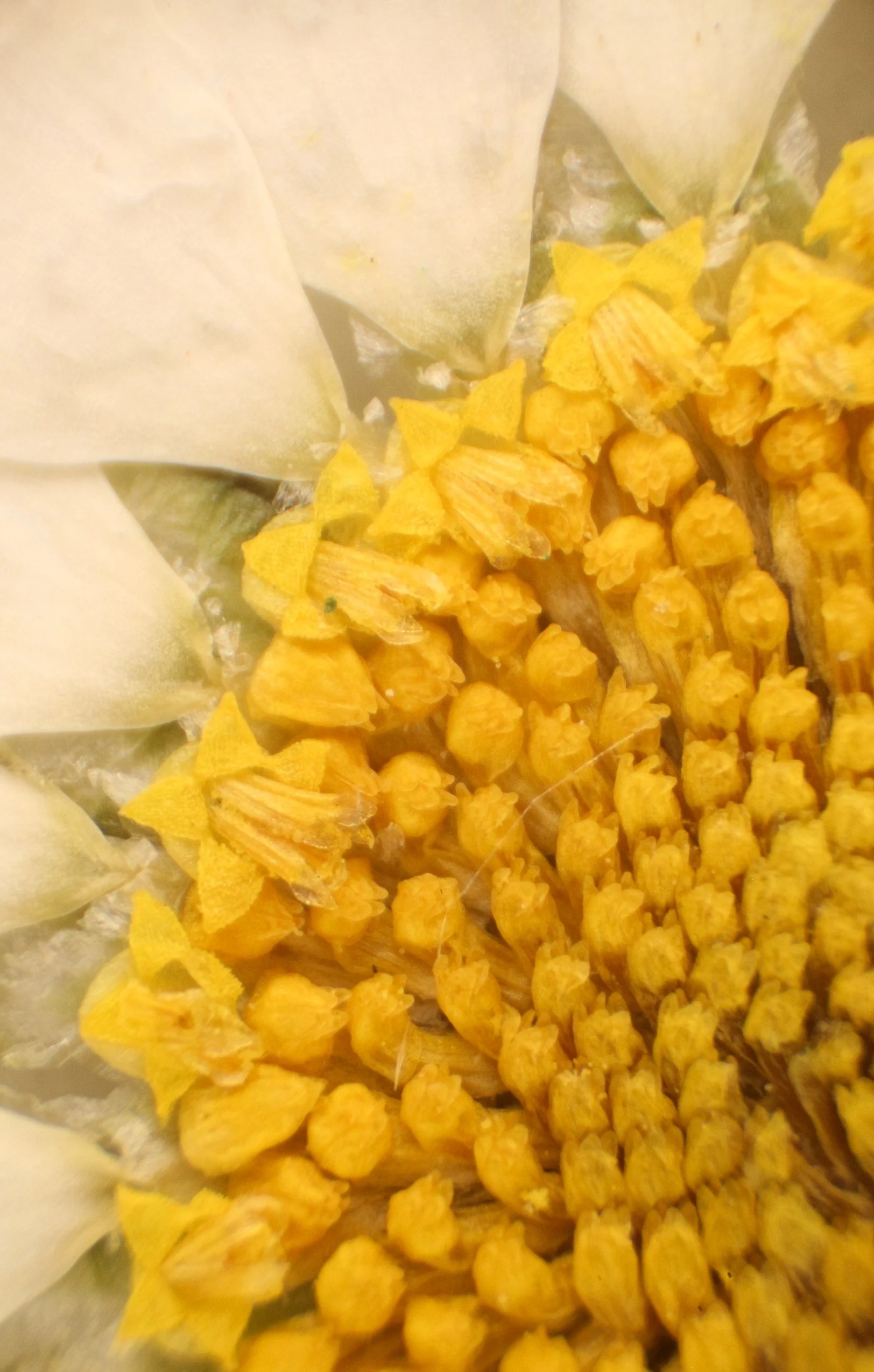
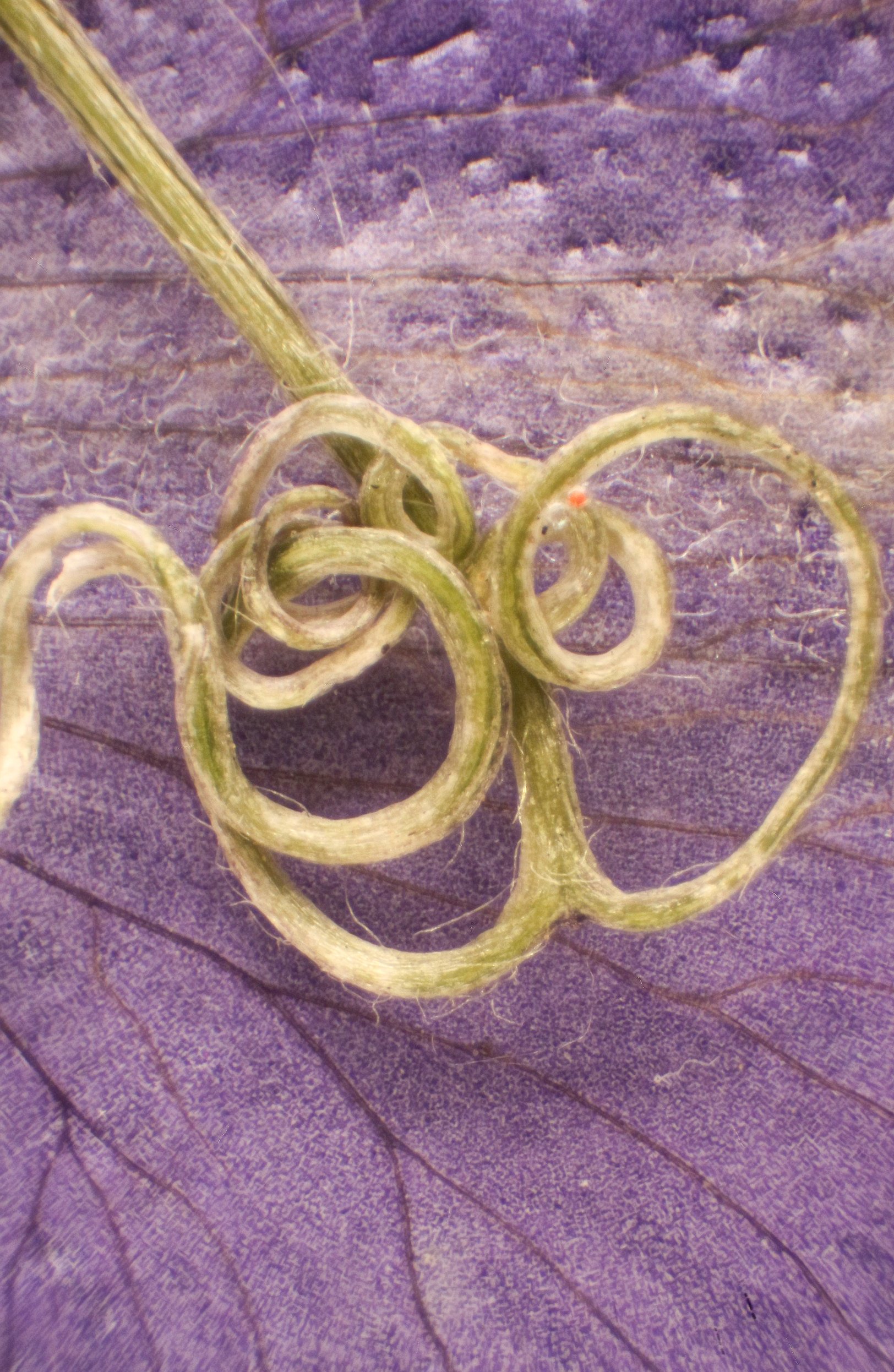
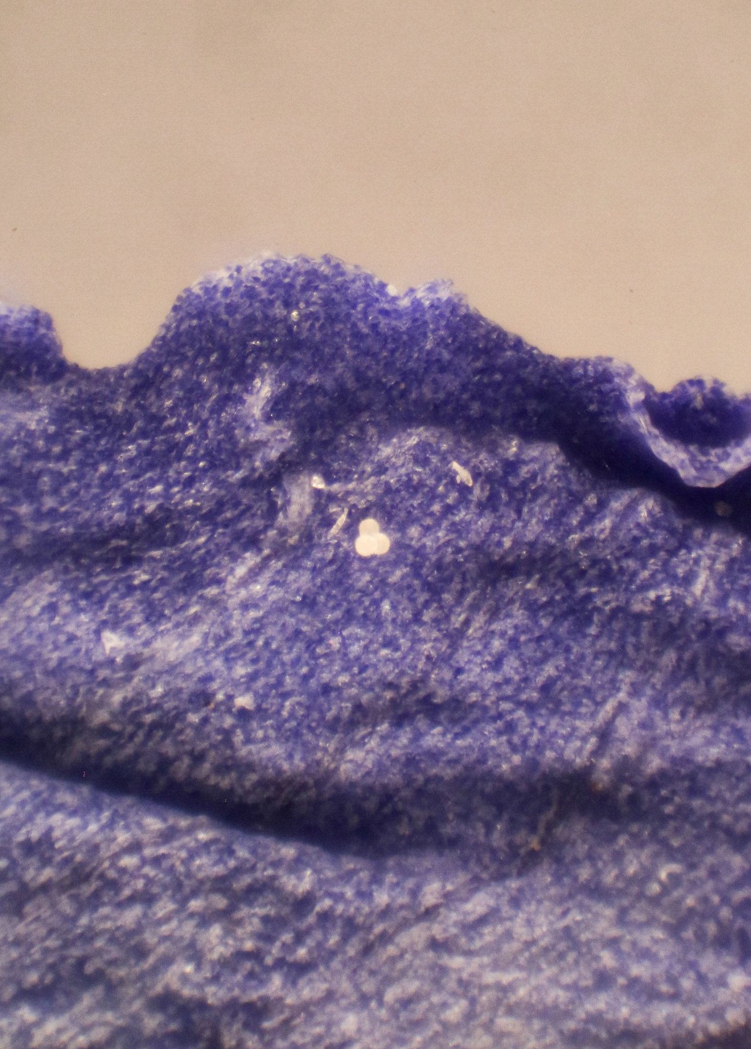
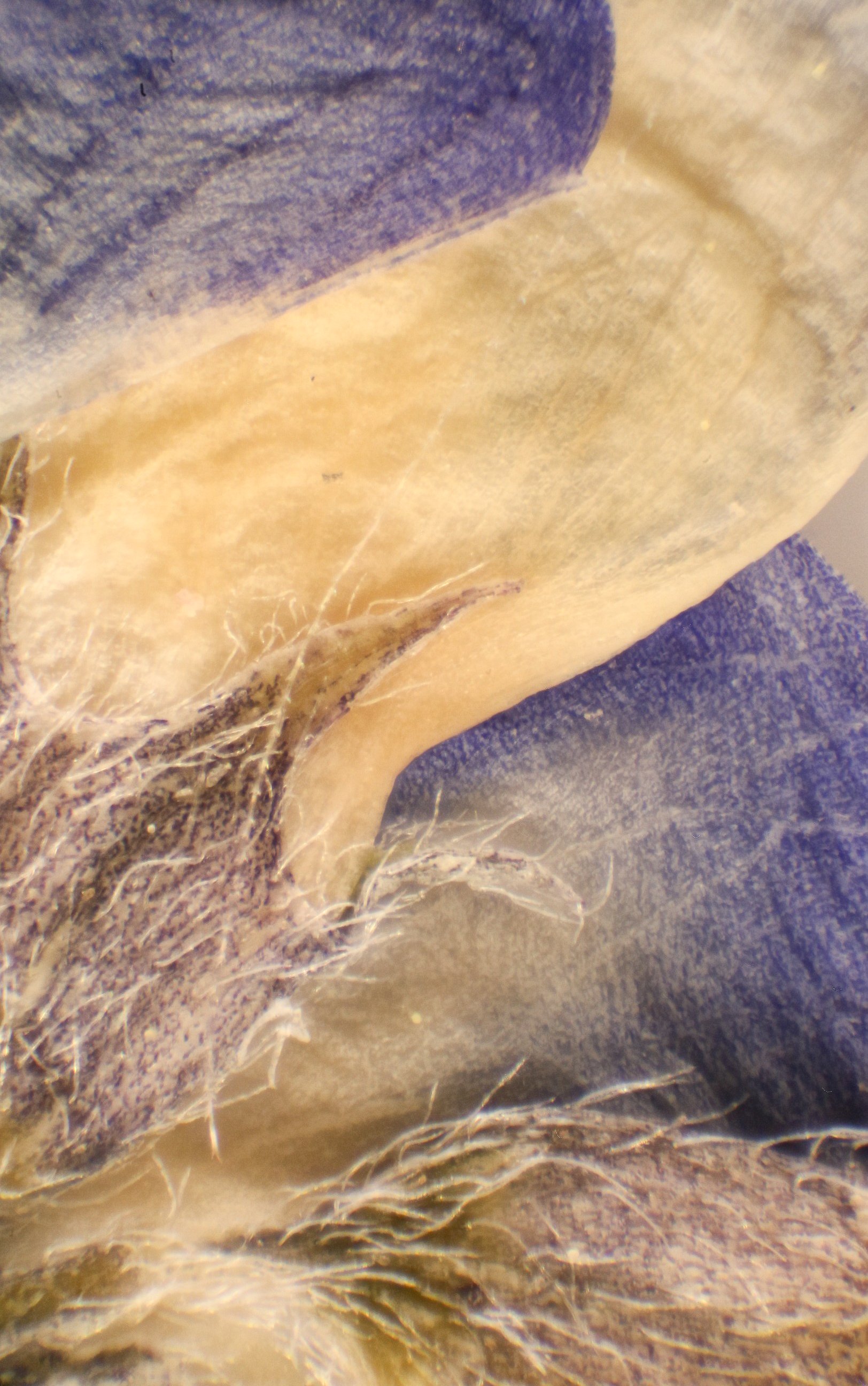
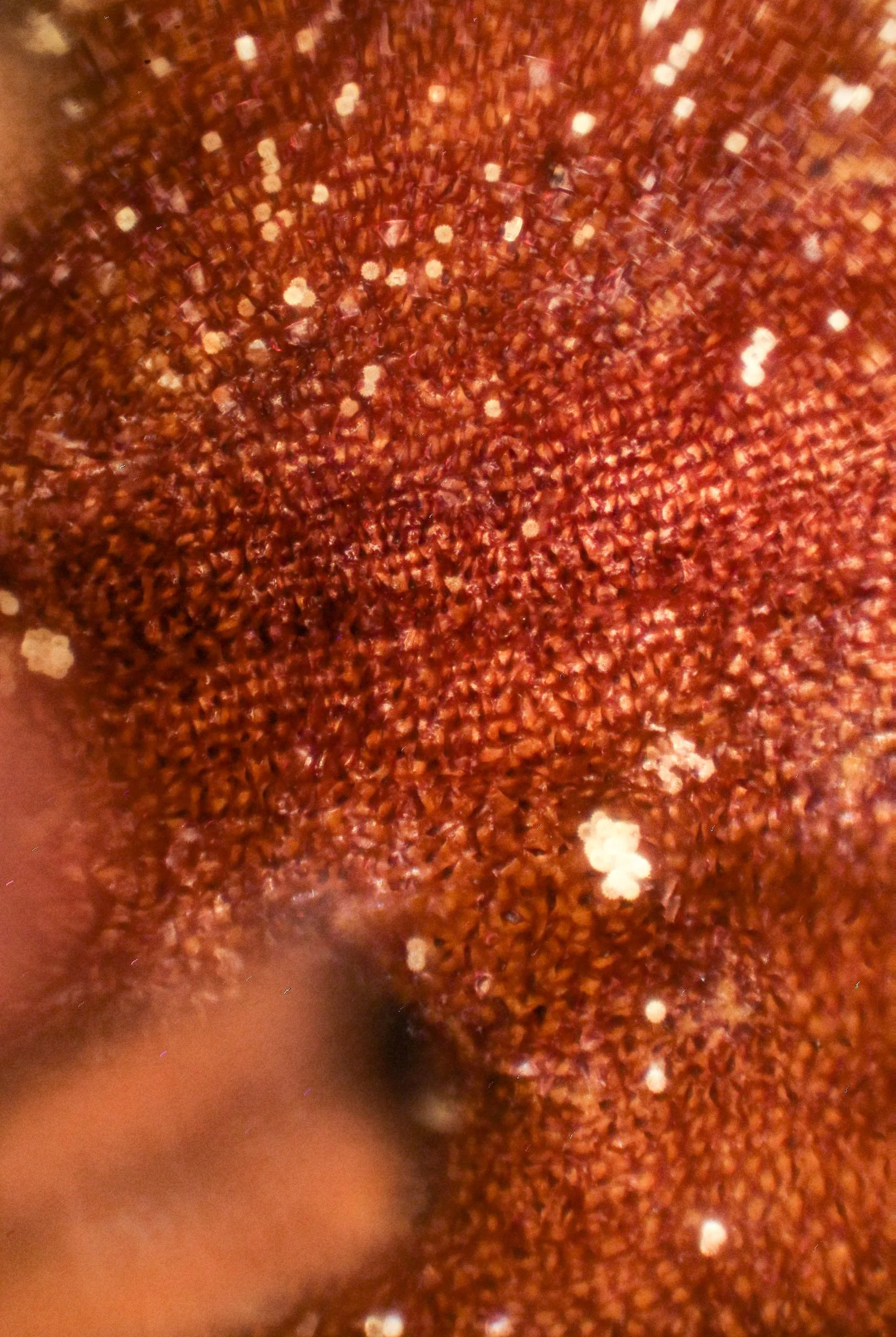
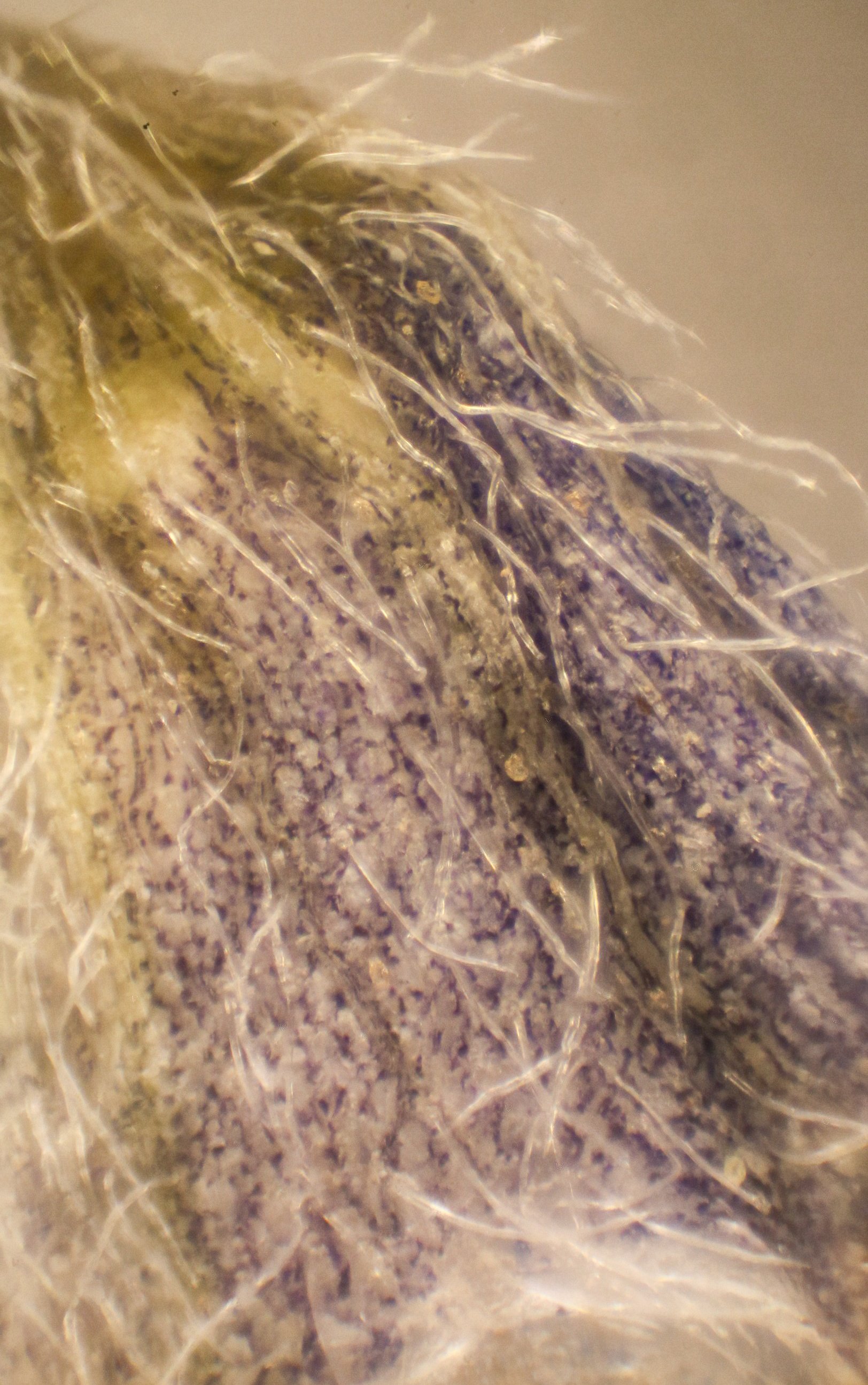
Flower reproductive systems under the microscope @ 4x, 8x, and 20x magnification.
Photomicrographs by Jehnifer Henderson.

